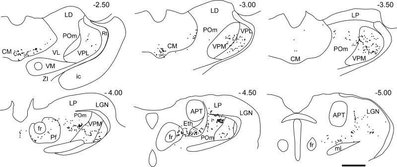
Fig. 5.
Plotted reconstructions of FG-labeled neurons in a series of coronal sections through the thalamus following the tracer injection depicted in Fig 2. Numbers in top right indicate the distance caudal from bregma. Scale bar: 1 mm. APT, anterior pretectal; eth, ethmoid; LD, lateral dorsal; LGN, lateral geniculate; LP, lateral posterior; ml, medial lemniscus; VM, ventral medial (VM); ZI, zona incerta.

