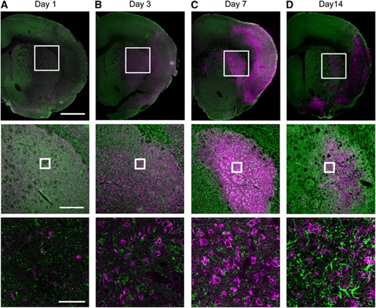
Figure 6.
Double-immunofluorescence staining of CD11b (magenta) and glial fibrillary acidic protein (green) in the transient middle cerebral artery occlusion (tMCAO) rat brain. Representative photographs showing brain sections in the ischemic core region at 1, 3, 7, and 14 days after tMCAO (A–D). Scale bars in the top, middle, and bottom row are 1 mm, 300 μm, and 50 μm, respectively.

