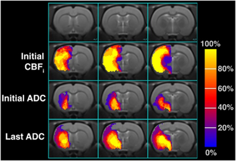
Figure 2.
Frequency of initial perfusion, initial (39±6 minutes), and late (237±54 minutes) diffusion lesions across eight rats in three coronal slices corresponding to bregma +0.7, −1.0, and −1.3 mm in the Paximos-Watson Rat Brain atlas. Voxels with values of 100% mean eight rats had a lesion voxel in that location. Voxels with values of 0% show that none of the eight rats exhibited a lesion at that spatial position. ADC, apparent diffusion coefficient; CBFi, cerebral blood flow index.

