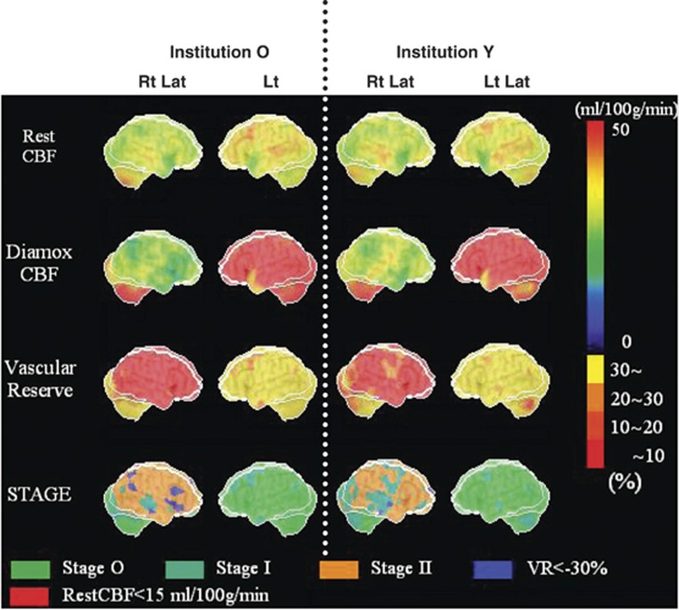
Figure 2.
Stereotactic extraction estimation based on the JET study (SEE-JET) images of patient 3 obtained in institutions O and Y. Rt Lat and Lt Lat indicate right hemisphere and left hemisphere outer lateral images, respectively. Cerebral blood flow at rest (Rest CBF), CBF after acetazolamide challenge (Diamox CBF), cerebrovascular reserve (vascular reserve), and severity of hemodynamic cerebral ischemia (STAGE) are shown as three-dimensional cerebral surface images. Images in institutions O and Y are visually almost identical.

