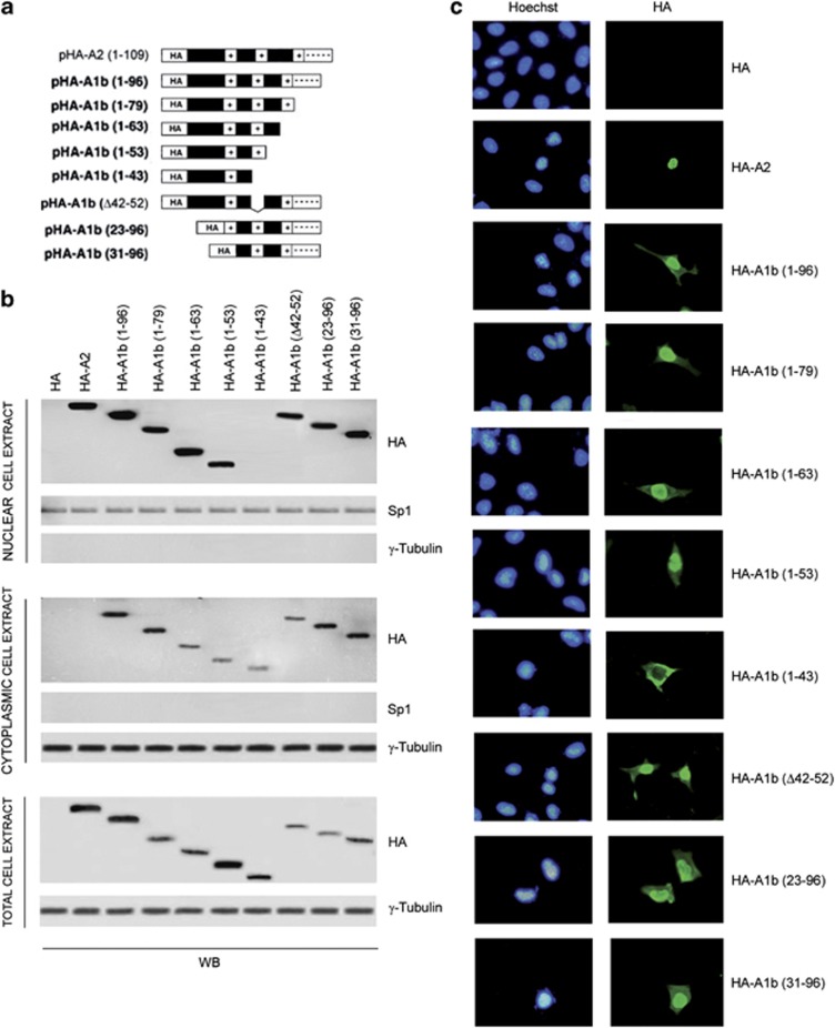
Figure 1.
HMGA1 is stably present at cytoplasmic level. (a) Diagram of the HA-tagged Hmga2 and wild-type and Hmga1 deletion mutants used in western blotting and immunofluorescence analysis. The AT-hook domains (+) and the acidic tail (- - - - -) are indicated. (b) Nuclear, cytoplasmic and total cell extracts from HEK293 cells were analyzed by western blotting for the expression of the constructs. Sp1 and γ-tubulin were used as markers of nuclear/cytoplasmic separation as well as loading controls. (c) Subcellular localization of HA-HMGA2 and HA-HMGA1 mutant proteins in HEK293 cells transfected with the indicated vectors. Nuclei were stained with Hoechst

