Figure 1.
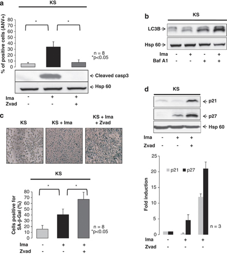
Imatinib-induced senescence of K562 cells is potentiated by caspase inhibition. K562 cells were grown in the presence of vehicle only, imatinib (Ima, 1 μM) or preincubated 30 min with Z-vad (50 μM) and then Ima (1 μM) for 48 h. An aliquot was incubated for 10 min in the presence of annexin-V-FITC. Samples were analyzed by flow cytometry and labeled cells were quantified as described in Materials and Methods. Results from eight experiments are expressed as the percentage of annexin V-labeled cells (a). Samples treated as above were used for detection of the cleaved form of caspase 3 by western blot and Hsp60 (as loading control). LC3B proteins were detected by western blot and Hsp60 was used as loading control (b). K562 cells treated as above (105 cells) was washed in PBS, fixed in PFA (4%) during 15 min and then incubated overnight in a 96-well plate in the presence of X-Gal (1 mg/ml) at 37 °C as described in Materials and Methods. The day after, the cells were washed once in PBS and SA-β-Gal activity was detected by a blue cell staining visualized under an inverted microscope. Pictures were acquired and analyzed using the NIS Nikon software. A representative result is shown in the top of the figure (c). SA-β-Gal-positive cells were quantified by counting 102 cells on three separate fields for each condition. Results show the mean of eight independent experiments (c). The proteins p21, p27 and Hsp60 were detected and the latter was used as a loading control. Results are from one experiment representative of three. The level of expression of p21 and p27 were quantified by densitometry analysis using ImageJ and results were normalized to the untreated condition (d)

