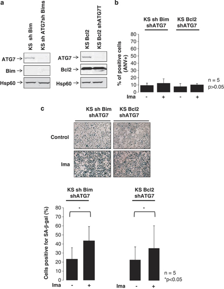
Figure 4.
Inhibition of apoptosis and autophagy limits imatinib-induced K562 cell senescence. K562 sh Bim and K562 Bcl2 cells were infected by incubation for 24 h with lentivirus coding for a shRNA anti-ATG7. Then, the cells were washed twice in PBS and grown for 6 days before sorting as described in Materials and Methods. Samples were analyzed for Bim, Bcl2 and ATG7 expression by western blotting (a). K562 sh Bim/sh ATG7 and K562 Bcl2/sh ATG7 cells were incubated with vehicle only or imatinib (Ima, 1 μM) for 48 h. Then, an aliquot was incubated for 10 min in the presence of annexin-V-APC. Samples were analyzed by flow cytometry and labeled cells were analyzed as described in Materials and Methods. Results from five experiments are expressed as the % of annexin V-labeled cells (b). K562 sh Bim/sh ATG7 and K562 Bcl2/sh ATG7 cells were treated as above (105 cells) and washed in PBS, fixed in PFA (4%) during 15 min and then incubated overnight in a 96-well plate in the presence of X-Gal (1 mg/ml) at 37 °C as described in Materials and Methods. The day after, the cells were washed once in PBS and SA-β-Gal activity was detected by a blue cell staining visualized under an inverted microscope. Pictures were acquired and analyzed using the NIS Nikon software. A representative result is shown in the top of the figure (c). SA-β-Gal-positive cells were quantified by counting 102 cells on three separate fields for each condition. Results show the mean of five independent experiments (c)

