
Fig. 4.
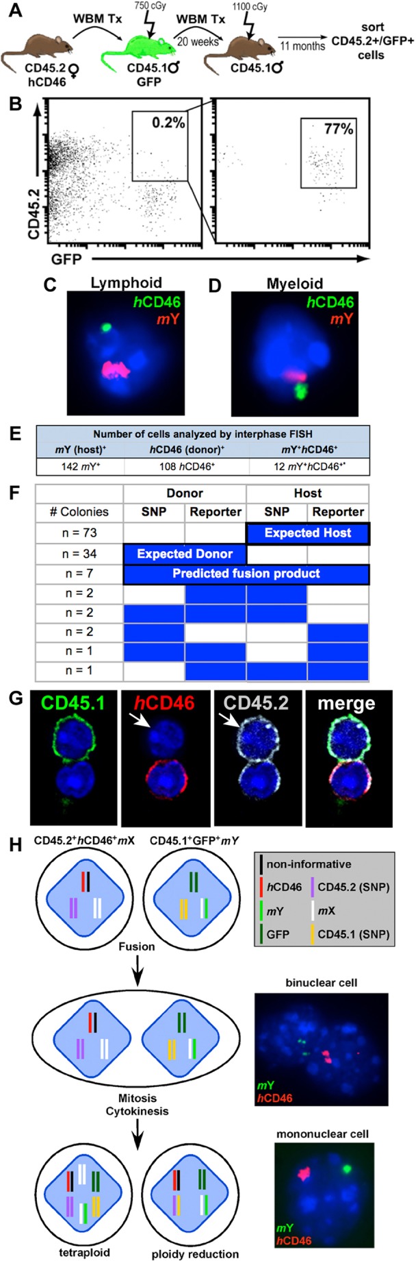
Fused cells contribute to long-term hematopoiesis in secondary recipients. (A) Secondary transplantation scheme. (B) Cells co-expressing CD45.2+ GFP+ were FACS sorted from spleens collected from secondary hosts. (C) Interphase FISH analysis of a fused lymphoid (CD4, CD8, B220) and (D) myeloid (Mac1, Gr1) cell isolated from spleen. Cells were probed for mouse Y (red) and human CD46 (green). (E) Hematopoietic cells co-expressing donor and host markers isolated from secondary transplant recipients analyzed by interphase FISH. (F) CFU-Cs were isolated from transplant recipients. SNP-PCR and PCR for autosomal reporter genes (CD46 or GFP) was performed on each colony. (G) Whole bone marrow (WBM) was harvested from a primary transplant recipient, FACS sorted for co-expression of parental markers and cytospun onto slides. Cells were stained with antibodies against host (CD45.1, FITC), donor (CD46, PE) and donor (CD45.2, APC). Although all CD46+ cells should be CD45.2+, there were a few instances in which a host marker was acquired and one of the two donor markers was lost. (H) Model of cell fusion. The genetic markers used in our studies are shown for each cell type. Fusion of the cellular membrane results in a binuclear cell containing a nucleus from each parental cell type. Following mitosis and cytokinesis, a daughter cell will be tetraploid or will undergo ploidy reduction to revert to diploid (or near-diploid) state with concurrent gain or loss of excess chromosomal material. In these cases, the daughter cell is mononuclear.