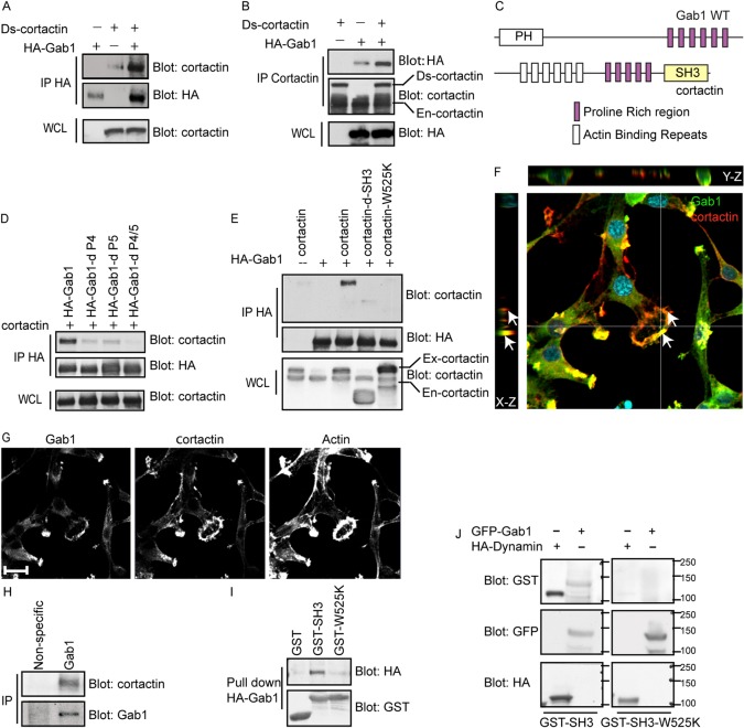
Fig. 7.
Cortactin interacts with Gab1 through its SH3 domain with proline-rich regions on Gab1. HEK 293 cells were transiently transfected with the indicated constructs; proteins from lysates were immunoprecipitated with HA antibody (A,D,E) or cortactin antibody (B) and immunoblotted as indicated. (C) Schematic diagram depicting a potential interaction of the cortactin SH3 domain and Gab1 proline-rich consensus motifs. (F,G) FR3T3 cells expressing Tpr-Met were transiently transfected with GFP-Gab1 and then plated on gelatin matrix for 24 hours. Cells were stained for GFP, cortactin and phalloidin. (F) X–Z and Y–Z projection of a 40 Z-stack showing the localization of Gab1 and cortactin to membrane protrusions (arrows). (H) Endogenous Gab1 was immunoprecipitated from BT549 cells and probed for cortactin. (I) GST–cortactin SH3 or GST–cortactin SH3-W525K mutant fusion proteins were coupled to GST beads and used to pull down proteins from lysates of HEK 293 cells transiently expressing HA–Gab1. (J) Proteins from HEK 293 cells, transfected with GFP-Gab1 or HA-dynamin, were immunoprecipitated with either HA or GFP antibody. The immune complex was separated by SDS-PAGE and transferred to a nitrocellulose membrane. Nitrocellulose membranes were incubated with fusion proteins of either cortactin GST–SH3 domain or GST–SH3-W525K and immunoblotted as indicated. Scale bars: 10 μm.

