
Fig. 1.
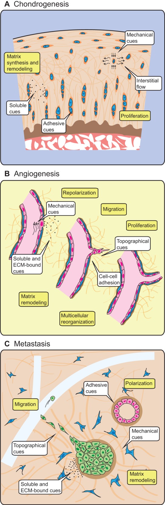
3D cellular phenomena in development, tissue homeostasis and disease are conducted by adhesive, mechanical and chemical cues originating from other cells and the extracellular environment. (A) Chondrocytes (blue) reside within a specialized pericellular ECM, where they are exposed to compressive forces, interstitial fluid flow, adhesive cues and soluble cues in the form of cytokines, which allow the cells to form and maintain the surrounding cartilage. (B) In response to soluble and matrix-bound growth factors and flow-induced mechanical forces on the blood vessel wall, endothelial cells (pink) alter their polarity and cell-cell contacts, and degrade the surrounding basement membrane (brown) and stromal ECM (orange) in order to collectively invade the surrounding tissue and form tubular sprouts. (C) The formation of normal epithelial structures (pink) requires adhesive and mechanical cues from neighboring cells and the basement membrane (brown) in order to tightly regulate proliferation and apoptosis. Misregulation of proliferation through genetic or extracellular changes initiates a cascade of soluble signals that activate fibroblasts (blue) in the surrounding stroma. Subsequent mechanical and structural changes in the stromal ECM enable transformed epithelial cells (green) to migrate towards neighboring vasculature (light blue) and, eventually, to metastasize. Drawings not to scale.