Fig. 3.

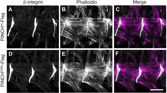
Muscle structure is preserved in PINCHQ38A-Flag rescued embryos. Stage 16–17 embryos were stained with anti-β-integrin antibody (A,C,D,F) and Phalloidin (B,C,E,F) to mark the muscle attachment site (green) and the actin cytoskeleton (magenta). β-integrin localizes normally to the muscle cell membrane and Phalloidin staining indicates that the actin cytoskeleton is stable and that the muscle fibers are properly distributed between muscle attachment sites in both PINCHwt-Flag rescued embryos (A–C) and in PINCHQ38A-Flag rescued embryos (D–F). Scale bar: 20 µm.
