Fig. 4.

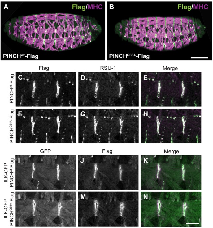
PINCH, RSU-1, and ILK are properly localized in PINCHQ38A-Flag rescued embryos. (A,B) Stage 16-17 embryos were stained with anti-Flag antibody to label PINCH transgenic protein (green) and anti-Myosin Heavy Chain (MHC) to label the embryonic body wall muscle (magenta). PINCHwt-Flag and PINCHQ38A-Flag localization and muscle structure are normal throughout the embryo. Scale bar: 50 µm. (C–H) Stage 16-17 embryos stained with anti-Flag antibody (green) to label PINCH transgenic protein (C,E,F,H) and anti-RSU-1 antibody (magenta) (D,E,G,H) show that both proteins localize in the absence of a PINCH-ILK interaction. Colocalization appears white in the merge (E,H). (I–N) Stage 16-17 embryos stained with anti-GFP antibody to label ILK-GFP (green) (I,K,L,N) and anti-Flag antibody to label PINCH transgenic protein (magenta) (J,K,M,N) show that PINCH and ILK colocalize in the absence of their direct interaction. Colocalization appears white in the merge (K,N). Scale bar: 20 µm.
