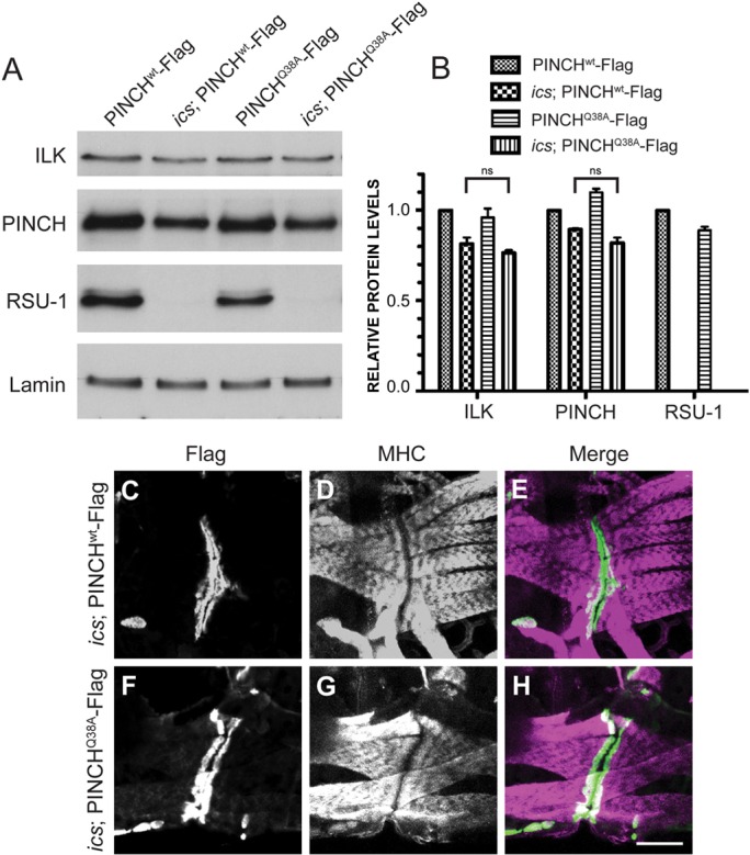
Fig. 7.

PINCH protein levels and localization are normal in ics; PINCHQ38A-Flag rescued larvae. (A) Western blots of pooled larvae at 66 hours AEL were probed with antibodies raised against ILK, PINCH, RSU-1 and Lamin. (B) Protein levels for the genotypes indicated were quantified and are represented as a fraction of PINCHwt-Flag expression. There is no statistical difference in PINCH or ILK levels between ics; PINCHwt-Flag and ics; PINCHQ38A-Flag rescued larvae. Bars represent the mean ± range for two replicate experiments. (C–H) 66 hour larval fillets were immunostained with anti-Flag antibody to label transgenic PINCH (green) (B,D,E,G) and anti-myosin heavy chain (MHC) to label the body wall muscle (magenta) (C,D,F,G). PINCH transgenes localize at muscle attachment sites in both ics; PINCHwt-Flag and ics; PINCHQ38A-Flag rescued larvae. Scale bar: 50 µm.
