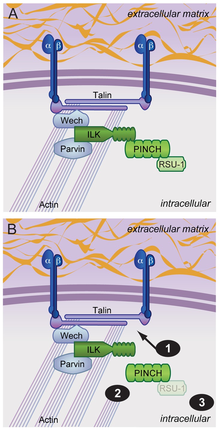
Fig. 8.

A model for ILK–PINCH–RSU-1 function. (A) Schematic of wild-type ILK, PINCH and RSU-1 within integrin adhesion complexes. A subset of protein interactions are highlighted demonstrating connection of the complex to integrins and the actin cytoskeleton. PINCH and ILK loss of function phenotypes are probably not due to the loss of a single protein, but to disruption of additional protein partners. (B) Specific disruption of the PINCH-ILK interaction does not affect viability or integrin function, and suggests that PINCH makes additional contacts that (1) anchor it to integrin rich adhesion sites and (2) connect it to the actin cytoskeleton. Loss of RSU-1 in PINCHQ38A rescued flies (3) results in lethality that is not due to mislocalization or destabilization of PINCH, suggesting a novel function for RSU-1.
