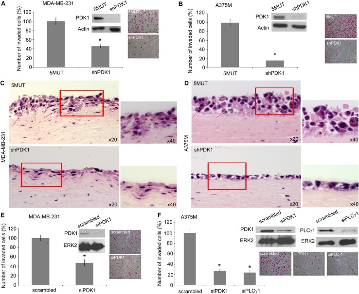
Fig. 6.
Inhibition of PDK1 blocks cancer cell invasion. (A,B) Results from an invasion assay of the indicated MDA-MB-231 and A375M stable cell populations on Matrigel. (C,D) Haematoxylin and Eosin staining of organotypic cultures of the indicated cells. Arrows indicate invading cells. Images are representative of two independent experiments for each cell line. Higher magnification views of the boxed regions are shown on the right of each image. (E) Results from invasion assay of MDA-MB-231 transiently transfected with siRNA targeting PDK1 or scrambled siRNA. (F) Results from invasion assay of A375M transiently transfected with scrambled siRNA or siRNAs targeting PDK1 or PLCγ1. Representative images of invading cells stained with Crystal Violet, and western blots confirming proteins downregulation, are also shown. All graphs show the number of invaded cells/field expressed as a percentage of control (5MUT or scrambled cells, respectively) and are means ± s.e.m. from three independent experiments. *P<0.05.

