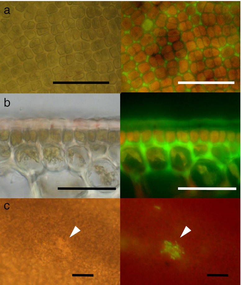
Fig. 4.
Bright field (left) and fluorescence (right) images of the vegetative region of Saccharina japonica sporophyte discs loaded with rhodamine 123. a Surface view of the vegetative region, showing the distribution of rhodamine 123 fluorescence in the interstitial area between cells. b Sectional view of the vegetative region, showing the fluorescence distributed in the cuticle, but mainly in the apoplast between the epidermal cells and outer cortical cells. c Surface view of the vegetative region with a small wounded region (arrows). Scale bars: 50 μm in (a) and (b) and 100 μm in (c)

