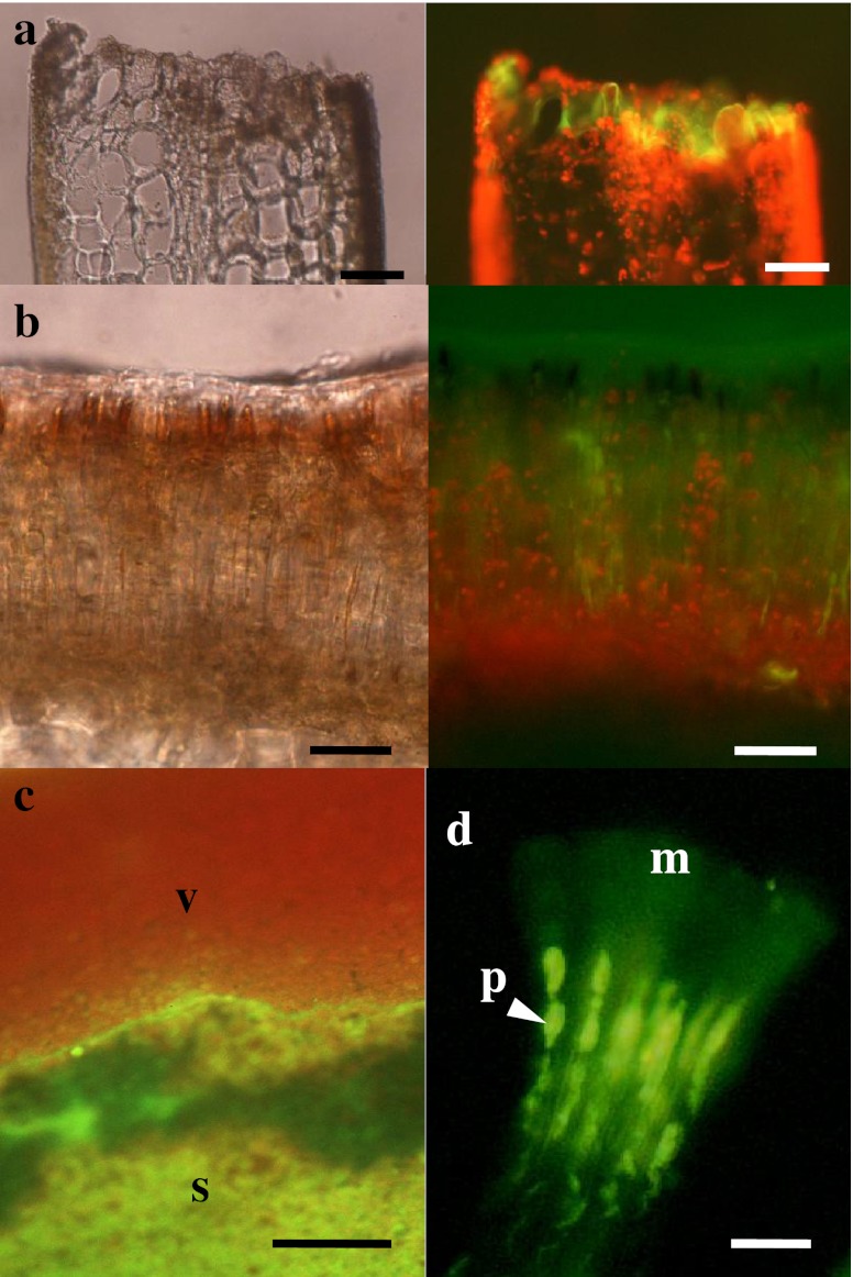
Fig. 5.
Bright field (left) and fluorescence (right) images of a wound (a) and sorus (b) in Saccharina japonica sporophyte discs loaded with rhodamine 123. (c) Fluorescent surface view image of a transitional area between the sorus (s) and non-sorus (V) portions, showing strong green fluorescence of rhodamine 123 in the sorus and red fluorescence of chlorophyll in the non-sorus portions. (d) Microscopic fluorescence image of separated paraphyses (p) with mucilaginous caps (m), showing the distribution of fluorescence in paraphyses and mucilaginous caps. Scale bars 50 μm in (a), (b), 100 μm in (c) and 10 μm in (d)

