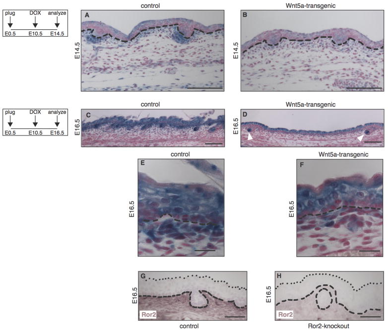
Figure 4. Loss of Wnt/β-catenin signaling at sites of Ror2 expression in the dermis of Wnt5a-overexpressing mice.
(A–F) Histological tissue sections of wholemount X-gal stained dorsal skin, demonstrating that expression of the Wnt/β-catenin reporter Axin2-lacZ is markedly reduced in Wnt5a-transgenic embryos at both E14.5 (A–B) and E16.5 (C–F). Mice depicted in A and B are littermates, as are the mice depicted in C and D. The activity of an independent Wnt/β-catenin reporter strain, TOPGAL, was also reduced at these timepoints (data not shown). (A–B) At E14.5 the overall activity of Axin2-lacZ is lower throughout the epidermis and dermis of Wnt5a overexpressing mice, but expression of the reporter is particularly reduced in the dermal condensates. (C–D) At E16.5 the Axin2-lacZ signal in the dermal papilla of existing hair follicles remains largely unaffected (white arrowheads in D). (E–F) Close-ups of the control and Wnt5a overexpressing skins shown in C and D, showing that the most dramatic reduction in Wnt/β-catenin reporter gene expression is seen in a thin layer of cells just underlying the basal layer of the epidermis in tetO-Wnt5a;R26rtTA double-heterozygous mice (F) but not in littermate controls (E).
(G–H) Immunohistochemical detection of endogenous Ror2 protein expression in the dermis of control (G) but not Ror2-knockout (H) skin at E16.5. Dashed lines indicate the boundary between epidermis and dermis. Dotted lines in G and H indicate the outermost cell layer of the epidermis. Scale bars are 100 μm in A–B, 20 μm in C and D, and 50 μm in E–H.

