Fig. 5.
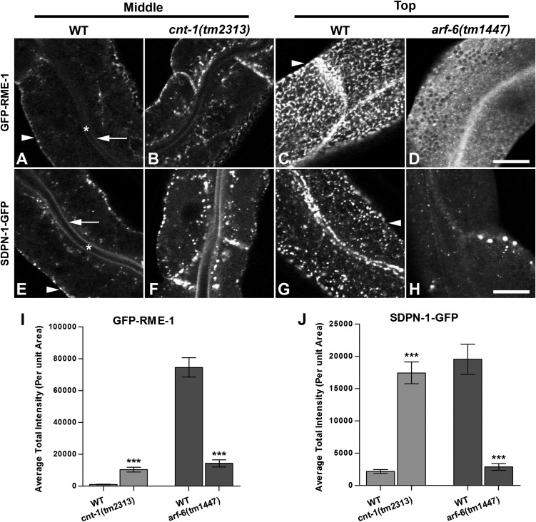
Aberrant subcellular distribution of PI(4,5)P2-binding proteins GFP-RME-1 and GFP-SDPN-1 in cnt-1 and arf-6 mutants. (A–D) GFP-RME-1 medial endosomal labeling increased in cnt-1 mutants, but its labeling of basolateral tubules and puncta decreased greatly in arf-6 mutants. (E–H) Similar to GFP-RME-1, recycling endosome marker GFP-SDPN-1 medial labeling increased in cnt-1 mutants, but labeling of basolateral tubules and puncta decreased greatly in arf-6 mutants. Asterisks in A and E indicate intestinal lumen, arrows in A and E indicate apical membrane, and arrowheads in A, C, E, and G indicate basolateral membrane. (I) Quantification of GFP-RME-1 average total intensity per unit area. (J) Quantification of GFP-SDPN-1 average total intensity per unit area. Error bars indicate SEM. n = 18; three different regions of the intestine (defined by a 100 × 100 pixel box positioned at random) were sampled in six animals of each genotype. ***P < 0.001, one-tailed Student’s t test. (Scale bar: 10 μm.)

