Fig. 3.
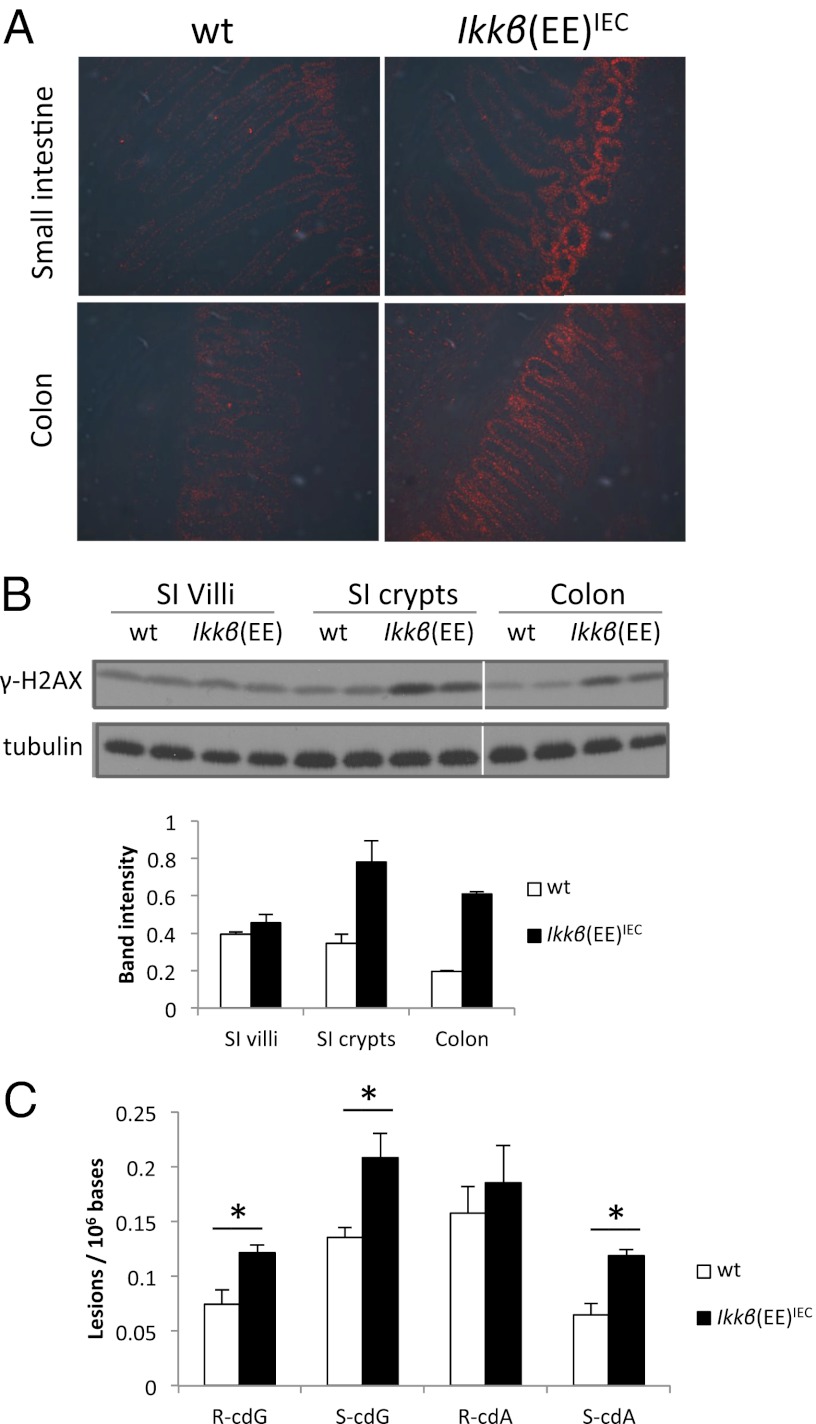
DNA damage in Ikkβ(EE)IEC mice. (A) Immunofluorescence analysis with anti–γ-H2AX antibody of frozen sections of intestinal tissue from 2-mo-old mice. (B) Immunoblot analysis γ-H2AX abundance in indicated intestinal regions of 2-mo-old mice. Shown are two mice per genotype with average band intensities indicated below. (C) Levels of 8,5′-cyclopurine-2′-deoxynucleosidesin mouse intestinal epithelium nuclear DNA (n = 3 for each genotype). cdG, 8,5′-cyclo-2′-deoxyguanosine; cdA, 8,5′-cyclo-2′-deoxyadenosine. R and S represent the 5′R and 5′S diastereoisomers, respectively.

