Fig. 1.
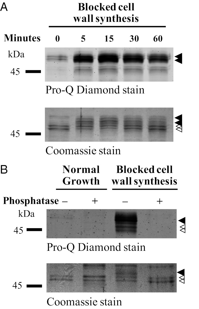
DivIVA is subject to phosphorylation. (A) Time course of DivIVA phosphorylation in response to the arrest of cell wall synthesis induced by bacitracin. Bacitracin (50 μg/mL) was added to growing cultures of WT S. coelicolor expressing FLAG-divIVA from a thiostrepton-inducible promoter. At the times indicated, cells were lysed, cell extracts were prepared, and FLAG-DivIVA/DivIVA was immunoprecipitated by using anti-FLAG affinity gel. (B) Phosphatase treatment of DivIVA. WT S. coelicolor expressing FLAG-divIVA was incubated with 50 μg/mL bacitracin for 60 min before harvest, preparation of cell extracts, and immunoprecipitation. The immunoprecipitated FLAG-DivIVA/DivIVA was analyzed before and after treatment with lambda protein phosphatase. Closed arrowheads indicate phosphorylated DivIVA and open arrowheads indicate nonphosphorylated DivIVA.

