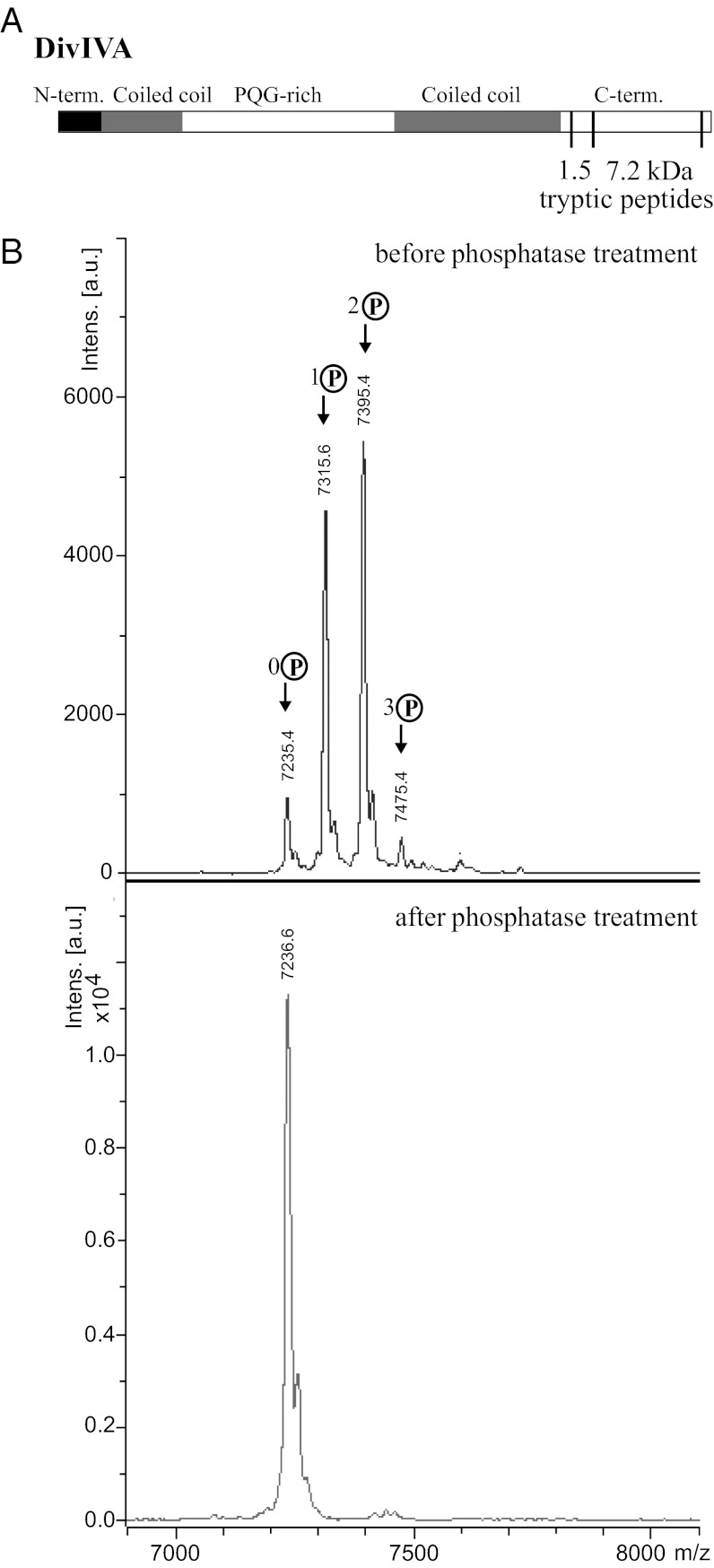
Fig. 2.
S. coelicolor DivIVA is multiply phosphorylated in the C-terminal region. (A) Schematic showing the positions within the DivIVA primary sequence of the 7.2-kDa phosphorylated peptide (residues 315–389) relative to the 1.5-kDa phosphorylated peptide (residues 301–314) described in the text. (B) Upper: MALDI mass spectrum of a 7.2-kDa tryptic fragment derived from the C-terminal region of DivIVA showing 0 to 3 phosphorylations (+80, +160, and +240 Da). Lower: Disappearance of the phosphorylated species upon treatment of the protein with calf intestinal alkaline phosphatase.

