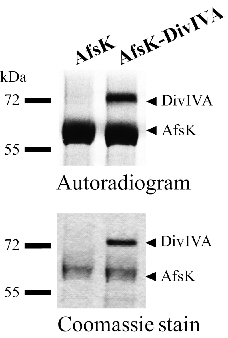
Fig. 4.
In vitro phosphorylation of DivIVA by AfsK. Recombinant AfsK and DivIVA were incubated with [γ-33P]ATP. Samples were separated by SDS/PAGE, visualized by autoradiography (Upper), and Coomassie-stained (Lower). Lower bands in the autoradiogram illustrate the autokinase activity of AfsK, whereas upper bands reflect DivIVA phosphorylation. In control experiments, DivIVA alone did not show any autophosphorylation activity.

