Fig. 5.
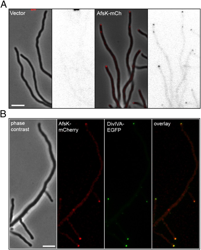
The DivIVA kinase AfsK localizes to hyphal tips. (A) S. coelicolor WT strain carrying empty vector pKF210 with promoterless mCherry (Left) or plasmid pKF255 expressing a translational afsK-mCherry fusion (Right). Representative images of growing hyphae are shown as phase-contrast image with overlaid fluorescence in red, and as the fluorescence image alone in inverted grayscale. (B) Colocalization of DivIVA and AfsK demonstrated by using an S. coelicolor strain producing both DivIVA-EGFP (green) and AfsK-mCherry (red). A series of images were collected for each channel, moving focus 0.2 μm between each image. The z-stacks were deconvolved by using Volocity software, and a central focal plane through the middle of the cells is shown as (from left to right) phase-contrast image, mCherry fluorescence, EGFP fluorescence image, and overlay of the fluorescence channels. (Scale bar, 4 μm.)

