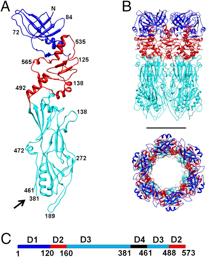
Fig. 4.
Structure of the major tail protein, gp12. (A) A monomer of gp12 with domains D1, D2 and D3 colored in blue, red and cyan, respectively. Black arrow points to the location of D4 where the protein was cleaved by trypsin. (B) Side and top views of the gp12 hexamer. Scale bar is 50 Å long. (C) Location of the domains on the linear sequence of gp12, using the same color scheme as in A and B.

