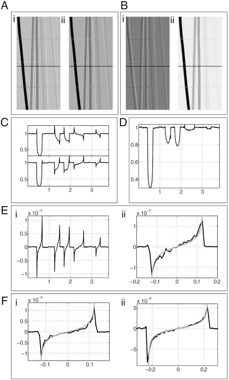
Fig. 2.
Phantom images and profiles. (A) Raw images, IR (i) and IL (ii) of a phantom of filaments made of titanium (125 μm radius), sapphire (125 μm radius), aluminium (125 μm radius), PEEK (225 μm radius), and PEEK (100 μm radius) respectively, going from left to right. (B) Extracted DP (i) and absorption (ii) images. (C) Raw image profiles along the line indicated in images A, i and ii. (D) Absorption image profile. Note how the raw image profiles for titanium in C possess only positive peaks and yet the peaks are still nulled in the absorption image. This is due to a subtle asymmetry in the raw profiles and is explained further in the SI Text. (E) DP image profile (i) and blow up of the titanium profile (ii) comparing the theoretical effective DP profile. (F) Monochromatic DP profiles of titanium (125 μm radius, i) and PEEK (225 μm radius, ii) along with theoretical DP profiles at the synchrotron photon energy of 20 keV. It should be noted that the plots in F are the only results in thus paper that were not acquired using a conventional source. The horizontal units of plots C–F are in millimeters.

