Fig. 1.
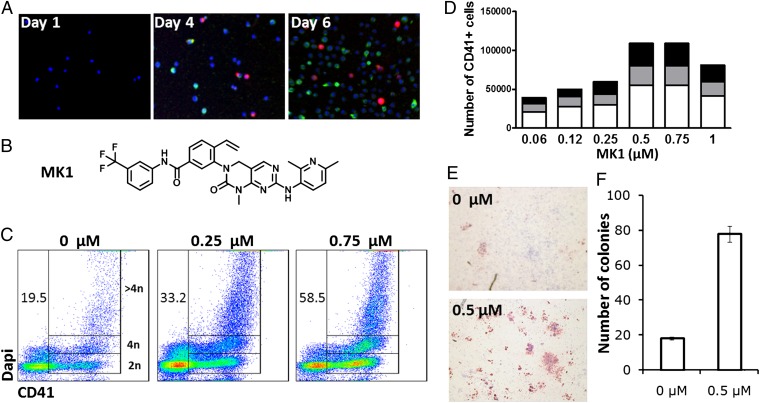
Screen of primary human mPB CD34+ cells identifies MK1 as an inducer of megakaryopoiesis. (A) Expression of CD41 (red) and CD71 (green) in mPB CD34+ cells cultured for 6 d in HSC expansion media. (B) Structure of MK1. (C) DNA content of mPB CD34+ cells cultured for 10 d in the presence of MK1 at the indicated concentrations. CD41+ cells containing 2N, 4N, and >4N are gated. Numbers represent the percentage of CD41+ cells. (D) Number of 2N CD41+ cells (white), 4N CD41+ cells (gray), and >4N CD41+ cells (black) from 5,000 mPB CD34+ cells cultured for 10 d in the presence of MK1 at the indicated concentrations. (E) CFU-Mk (red) generated from mPB CD34+ cells cultured in the presence or absence of MK1 (0.5 μM) for 8 d (F) Total CFU-Mk present in 5,000 mPB CD34+ cells expanded for 8 d in the presence or absence of MK1 (0.5 μM). Mean and SD are plotted from three individual plates within the same experiment (P < 0.01). Data are representative of a least three independent experiments.

