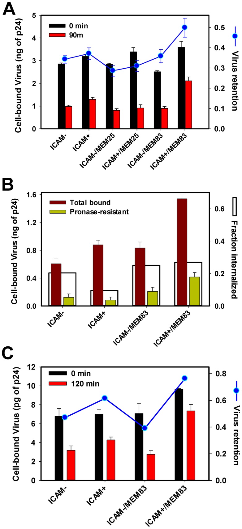
Figure 3. Effect of ICAM-1 on virus attachment to cells aided by centrifugation in the cold.
PM-1 cells (1·105) were adhered to poly-lysine-coated 96-well plates. Immobilized cells were pre-treated with MEM25 or MEM83 antibodies or left untreated. Eight ng of either ICAM+ or ICAM- viruses were added to cells and centrifuged at 4°C for 30 min. (A) After virus binding by spinoculation, cells were washed, and the amount of p24 was measured either immediately (0 min) or after incubation at 37°C for 1.5 h (90 min). Blue symbols show the ratio between the cell-associated virus after incubation at 37°C and immediately after spinoculation. (B) To determine the fraction of internalized viruses following the pre-binding step in the cold and incubation for 1.5 h at 37°C, cells were treated with 2 mg/ml pronase for 30 min on ice or left untreated. Cells were washed, and the remaining p24 was measured by ELISA. White bars are obtained by normalizing the pronase-resistant p24 signal to the amount of virus associated with untreated cells. (C) Same as in panel A, but using HIV-1 TYBE pseudoviruses. After ICAM− or ICAM+ viruses (1 ng of p24) were spinoculated onto cells in the cold, cells were washed and the amount of p24 was measured either immediately (0 min) or after incubation at 37°C for 2 h (120 min). Blue circles show the ratio between the cell-associated virus after incubation at 37°C and immediately after spinoculation. Data are means and SEM from a representative experiment performed in triplicate.

