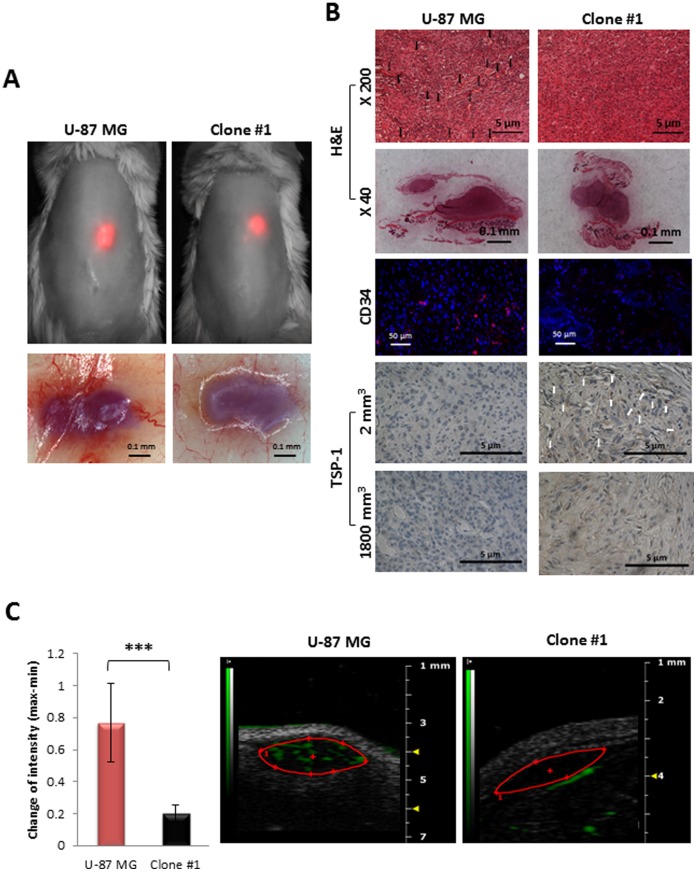
Figure 4. Comparison of size-matched tumors generated from U-87 MG and Clone #1 cells.
A. Sixteen days post subcutaneous inoculation of mCherry labeled U-87 MG and Clone #1 cells, both tumor types were approximately 2 mm3 in diameter and detectable by non-invasive CRI Maestro™ imaging system (upper panel). Flipped skin of tumor-bearing mice revealed highly vascularized U-87 MG-generated tumors, while blood vessels were only detectable in the surrounding skin of Clone #1-generated tumors (lower panel). B. H&E, CD34 (merged image. The separate images are provided as Fig. S2) and TSP-1 staining of U-87 MG and Clone #1 tumor sections. TSP-1 staining was done on size-matched tumors from day 16 (2 mm3) and on large tumors (U-87 MG tumors at end point of experiment and Clone #1 tumors after escape from dormancy) (1800 mm3). C. Contrast-enhanced US imaging of U-87 MG and Clone #1 subcutaneous tumors show high vascularization of the U-87 MG fast-growing tumor (red bar, n = 5) compared with Clone #1 dormant tumors (black bar, n = 3) (p = 0.008). Data represent mean ± s.d. *** p<0.01.

