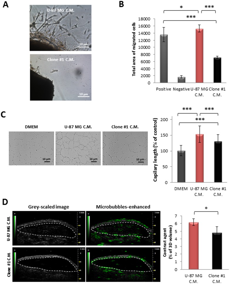
Figure 7. Angiogenic potential comparison between cells of U-87 MG and Clone #1.
A. C.M. of U-87 MG cells induced extensive sprouting of endothelial cells (upper panel) from aortic rings resected from mice as compared with C.M. of Clone #1 (lower panel). B. HUVEC migrate towards C.M. of U-87 MG cells (red bar) at a significantly increased rate (p = 1.6×10⁻¹³) compared with that of Clone #1 (black bar). DMEM containing 10% FBS served as positive control; DMEM served as negative control. C. HUVEC’s ability to form capillary-like tube structures on Matrigel® is significantly higher (p = 0.001) in the presence of C.M. of U-87 MG cells (red bar) compared with that in the presence of C.M. of Clone #1 cells (black bar). D. Contrast-enhanced ultrasound imaging of subcutaneously-inoculated plugs containing C.M. media from U-87 MG cells (red bar, n = 3) showed increased vascularization compared with C.M. from Clone #1 cells (black bar, n = 3) (p = 0.04). Data represent mean± s.d. from three independent experiments. * p<0.05, *** p<0.01.

