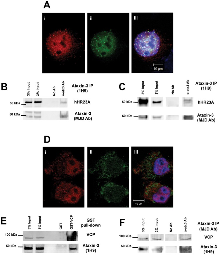
Figure 1. Ataxin-3 interacts directly with hHR23A and VCP/p97.
(A) Confocal microscopy images of COS-7 cells fixed and immunostained for endogenous ataxin-3 (i-red) and hHR23A (ii-green). DNA was stained with Hoescht 33342 (blue) in the merged image (iii). (B) Endogenous ataxin-3 was immunoprecipitated from COS-7 cell extracts and co-immunoprecipitation of endogenous hHR23A was determined by western blotting. (C) Recombinant human ataxin-3 and recombinant human hHR23A were incubated under the same conditions applied in the in vitro protease assays, for 5 hours at 37°C. Recombinant ataxin-3 was precipitated using anti-ataxin-3 (1H9) antibody and hHR23A co-immunoprecipitation was analyzed by western blotting. (D) Visualization of endogenous ataxin-3 (i-red) and VCP/p97 (ii-green) through confocal microscopy in fixed COS-7 cells using specific antibodies. Nuclei were stained Hoescht 33342 dye (blue) for merged images. (E) Recombinant GST-VCP/p97 fusion protein was used to pull-down endogenous ataxin-3 from COS-7 total extracts. Endogenous ataxin-3 and recombinant VCP/p97 were detected by western blotting. (F) Recombinant human VCP/p97 and recombinant human (Q22) ataxin-3 were added to a buffered solution at 37°C for 5 hours. These samples were used to immunoprecipitate ataxin-3 and co-immunoprecipitation of VCP/p97 was assessed by western blotting.

