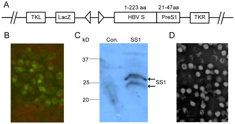
Figure 1. Characterization of recombinant vaccinia virus RVJSS1 and HBV particle-like subunit vaccine HBVSS1.
(A) Schematic diagram of recombinant vaccinia virus RVJSS1, which contains two expression cassettes in a back-to-back orientation, flanked by vaccinia virus TK region sequences. Virus RVJSS1 was a Tiantan strain vaccinia virus with a lacZ gene led by a p11 promoter inserted into the TK region; the expression cassette on the right consists of the SS1 fusion protein led by the 7.5 K promoter. (B) Immunofluorescence assay to confirm SS1 fusion protein expression. CEF cells were infected by the purified RVJSS1 virus and fixed, permeabilised, stained with rabbit anti-PreS1 antibody and fluorescein isothiocyanate (FITC) conjugated to a secondary antibody, and then visualised using fluorescence microscopy. (C) Western blot analyses to detect expression of SS1 fusion proteins in CEFs infected with RVJSS1 using specific antibodies. The expression bands of the SS1 proteins are indicated by arrowheads. (D) Negative staining of purified HBVSS1 particles vaccine using electron microscopy.

