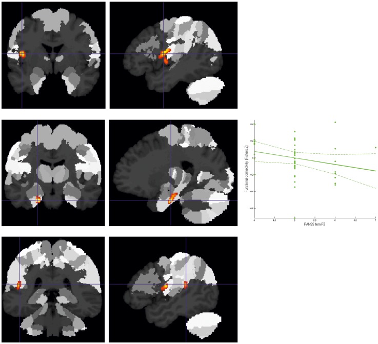
Figure 3. Decreased connectivity to lSTG in patients as compared to controls.
A. Cluster of significantly (p<0.05 corrected) decreased connectivity with the lSTG in patients in the left frontal operculum/anterior insula shown on the MPM of the SPM Anatomy Toolbox. B. Cluster of significantly (p<0.05 corrected) decreased connectivity with the lSTG in patients in the left hippocampus (subiculum/entorhinal cortex) shown on the MPM of the SPM Anatomy Toolbox. C. Cluster of decreased connectivity (as a strong statistical trend) with the lSTG in patients in the left temporo-parietal operculum/retroinsular cortex.

