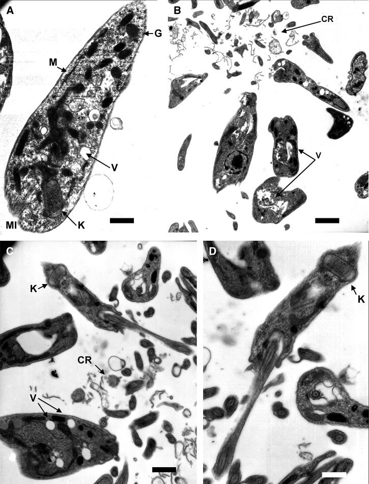
Figure 5.
Ultrastructural alterations by transmission electron microscopy (TEM) in Trypanosoma cruzi treated with terpenoids compounds. (A) Control parasite of T. cruzi with structures as vacuoles (V) and mitochondrion (M), kinetoplast (K), glycosomes (G), and microtubules (MI) (Bar: 583 μm). (B) Epimastigotes of T. cruzi treated with Compound 1 with cellular debris from dead parasites (CR) and vacuoles (V) (Bar: 1.59 μm). (C and D) Epimastigotes of T. cruzi treated with Compound 2 with cellular rest (CR), vacuoles (V), and swelling kinetoplast (K) (Bar: 1.00 μm and Bar: 583 μm, respectively).

