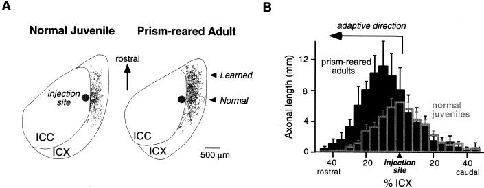
Figure 3.
Anatomical traces of learning in the ICX of the barn owl. (A) Digital image drawings of labeled axons in 40-μm thick, horizontal sections through the optic lobe of a normal juvenile (Left) and a prism-reared adult (Right). To visualize the pattern of axonal projection from the ICC to the ICX, the anterograde tracer, biocytin, was injected into the ICC at a site representing 20 μs ITD contralateral ear leading. The location of the injection site in the ICC is indicated as a dark circle. Labeled axons in the ICX are shown as thin lines. In a normal juvenile, the axonal projection field in the ICX is spatially restricted, symmetric, and centered around the rostral-caudal level of the injection site. In contrast, in a prism-reared adult expressing a rostral map shift, there is a dramatic expansion of the projection field in the rostral portion of the ICX. The direction of this axonal expansion is adaptive for the direction of prismatic displacement. Thus, these axons represent the learned projection field. (B) Composite spatial distributions of axons for normal juveniles (empty gray bars) and prism-reared adults (solid black bars). To quantify the spatial distribution for each case, the ICX was subdivided into measurement zones oriented orthogonal to the rostral-caudal axis. Each measurement zone corresponded to 5% of the rostral-caudal extent of the ICX. Total axonal length in each measurement zone was determined by computer analysis of the digital image drawings. Histograms were aligned relative to the rostral-caudal level of the injection site. Composite distributions were obtained by averaging the individual cases. In prism-reared adults with rostral map shifts (n = 4), the axonal density on the rostral flank of the distribution is significantly greater than in normal juveniles (n = 7; ANOVA, P < 0.0001), indicating that remodeling occurs by a net elaboration of axons. In contrast, the axonal density within the normal projection field, located at the rostral-caudal level of the injection site, is not significantly changed by prism experience. Data are from W.M.D., D. Feldman, and E.I.K., unpublished data.

