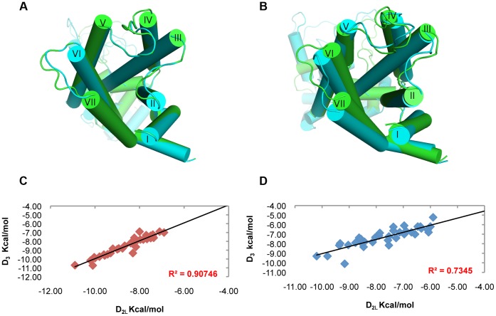
Figure 3. Structure differentiation of hD3 and hD2L receptors simulated in membrane.
(A) Superimposition of hD3 (green cartoon) and hD2 (cyan cartoon) homology models before the refinement with simulation in membrane. (B) structural alignment of hD3 (green cartoon) and hD2 (cyan cartoon) receptors after 3 ns of MD simulation in membrane. (C) high correlation of hD3 and hD2 binding energies (Autodock Vina) of D2-like ligands from homology models without MD refinement. (D) low correlation of hD3 and hD2 binding energies (Autodock Vina) of D2-like ligands after MD refinement.

