
Fig. 3.
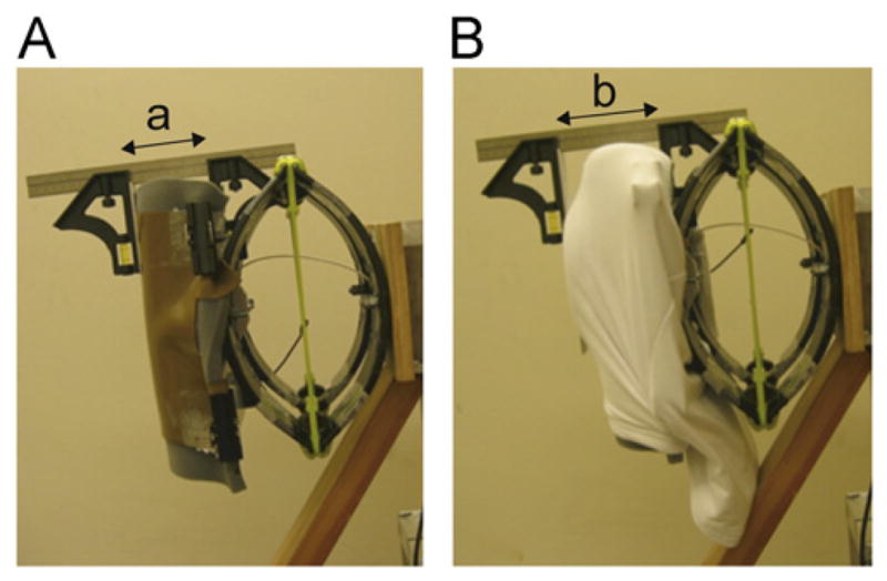
Illustration of the protocol for measuring the increase in pelvic width in the frontal plane when wearing the protector (thickwearing). This variable was calculated as the distance from the medial aspect of the surrogate pelvis to the lateral surface of the hip protector (measure ‘b’ in subplot B) minus the distance between the medial and lateral aspects of the unpadded surrogate pelvis (measure ‘a’ in subplot A).