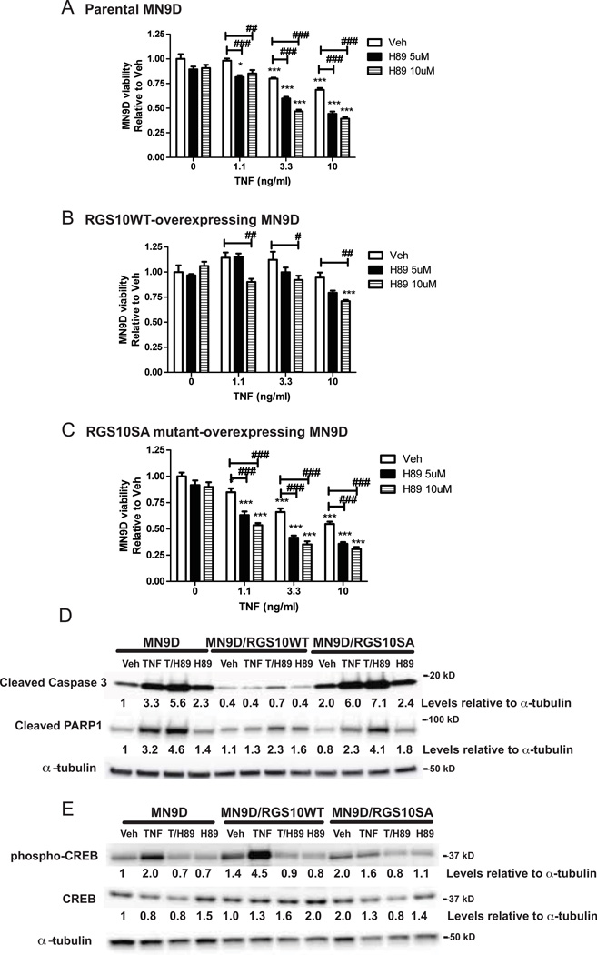
Figure 4. Neuroprotective effect of RGS10 in DA cells requires activation of cAMP-dependent protein kinase (PKA).
MN9D cells were neurally differentiated then treated with TNF (1.1, 3.3 or 10 ng/mL) for 48 hrs in the presence or absence of pre-treatment of H89 (5 or 10 µM) for 45 min. Cell viability was assessed using the MTS assay on A, parental MN9D viability, B, MN9D cells stably over-expressing WT RGS10 viability, and C, MN9D cells stably over-expressing RGS10 SA mutant. Values represent mean ± SEM. One way ANOVA followed by Tukey’s post hoc test, *, ** or *** denote significant differences between vehicle and TNF treatments within the group at p < 0.05, p < 0.01 or p < 0.001 respectively. #, ## or ### denote significant differences between the H89 treatments groups at p < 0.05, p < 0.01 or p < 0.001 respectively. Values shown are group means (n=6) ± S.E.M from one experiment representative of three independent experiments. D, Effect of PKA inhibition on TNF-induced caspase 3 and PARP1 cleavage by western blot analysis. Parental MN9D cells or MN9D cell lines stably over-expressing WT RGS10 or RGS10SA mutant were neurally differentiated then treated with TNF (10 ng/mL) in the presence or absence of H89 (10 µM). E, Effect of PKA inhibition on TNF-induced phospho-CREB and CREB levels by western blot analysis. Cells were prepared as described above. Densitometry and normalization to α-tubulin as a protein loading control was performed. All the western blots are representative of three independent experiments.

