Figure 3.

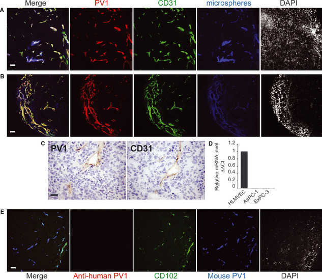
In both AsPC‐1 and BxPc‐3 pancreatic tumours PV1 is expressed only in tumour endothelial cells (A–B) Confocal microscopy of frozen sections from AsPC‐1 pancreatic tumours grown in nude mice, with anti‐PV1 (red) and anti‐CD31 (green) and antibodies directly coupled to fluorophores. The vascular lumen (blue) was labelled by the introduction of dark red microspheres in the circulation. In the merged field, white means triple co‐localization. Last panel in each shows nuclei stained with DAPI. (A) is a representative field showing vessels inside the tumour whereas (B) shows vessels at the tumour periphery. Magnification 20×. Bars – 20 μm. (C) Immunohistochemistry of serial frozen sections from human pancreatic xenograft tumours grown in nude mice, with anti‐PV1 (PV1) and anti‐CD31 (CD31) antibodies. The nuclei are counter stained with haematoxylin. Bars – 20 μm. (D) PV1‐mRNA levels in HLMVEC and human pancreatic tumour cell lines AsPC‐1 and BxPC‐3 measured by real‐time quantitative PCR. The data were obtained from triplicate samples and normalized to β‐actin levels (ΔΔCt). Bars – SEM (E) Confocal microscopy of frozen sections from AsPC‐1 pancreatic tumours grown in nude mice, stained with anti‐human PV1 (red) showing lack of expression of PV1 by human cancer cells. In the same specimen, mouse PV1 (blue) is present and colocalizes with the vascular marker CD102 (green), both detected by antibodies directly coupled to fluorophores. Magnification 20×. Bars – 10 μm.
