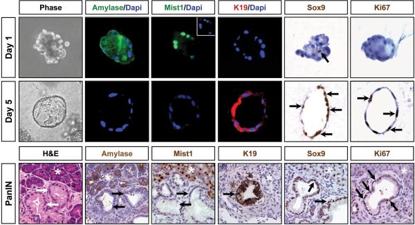
Figure 2. KrasG12D-expressing acinar cells undergo conversion to a ductal phenotype when maintained in 3D collagen matrix.
Fluorescence and light microscopy reveals that KrasG12D acinar cells express acinar gene products at day 1 but rapidly convert to a ductal cell phenotype with activation of K19 and Sox9 expression. Inset for day 1 Mist1 panel shows the Dapi stained nuclei for this acinus. Note that a single centroacinar cell (arrow) in an isolated acinus is SOX9 positive. Pancreas serial sections from experimental littermates confirm the absence of AMYLASE and MIST1 and the expression of SOX9 and K19 in acinar-derived PanINs (arrows). Both PanINs and ductal cysts are also Ki67 positive (arrows). asterisks - adjacent acinar tissue.

