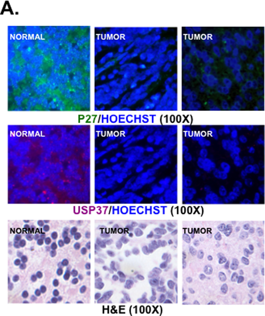
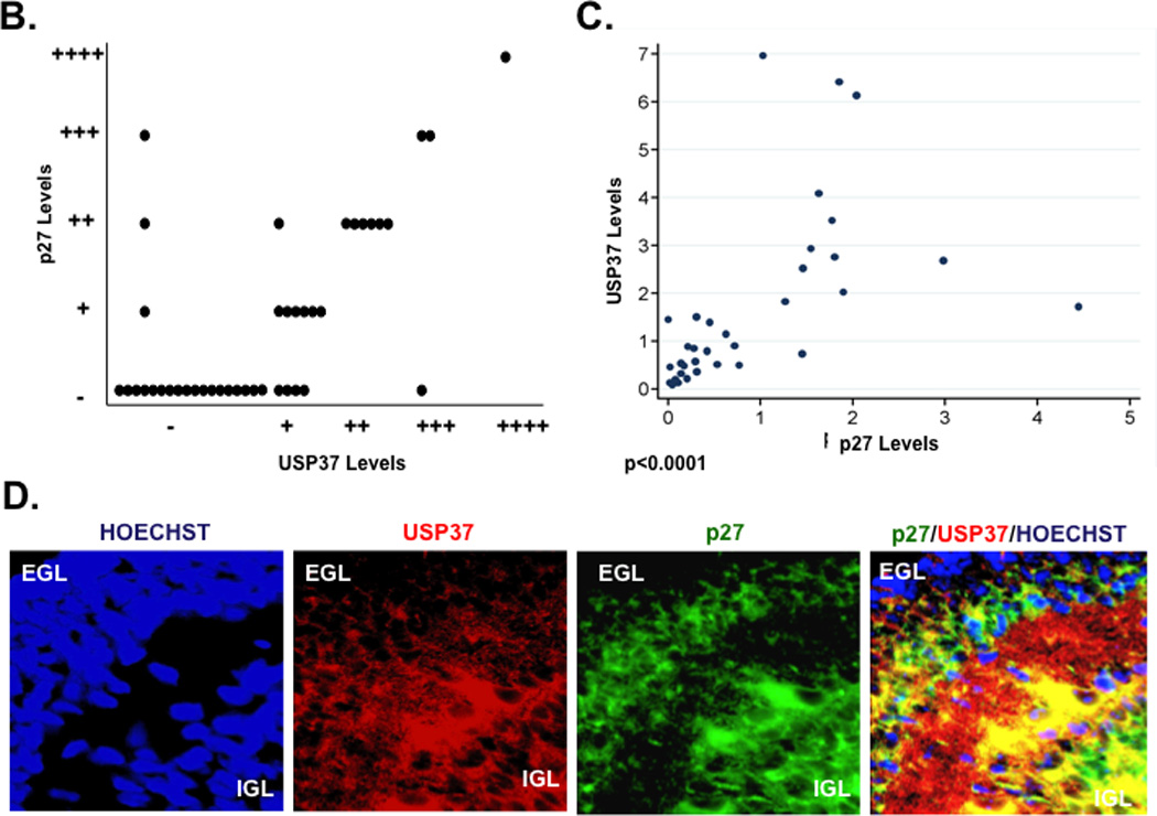
Figure 4. USP37 and p27 levels are correlated in human medulloblastoma and normal mouse cerebellum.


(A) Expression of USP37 (red) and p27 (green) in human medulloblastoma samples was assessed by IFA using specific antibodies and Cy3- or Alexa-488-conjugated secondary antibodies. Nuclei were stained with Hoechst dye and the images viewed and analyzed by fluorescence microscopy. (B and C) Distribution of p27 expression in USP37-expressing human medulloblastomas is provided. Significance and correlation were measured using the Wilcoxon rank-sum test and Spearman correlation test (rank correlation=0.67 and p<0.0001). (D) USP37 (red) and p27 (green) expression in cerebella of postnatal day 7 mice were evaluated by IFA as described above. EGL: external granule layer, IGL: Internal granule layer.
