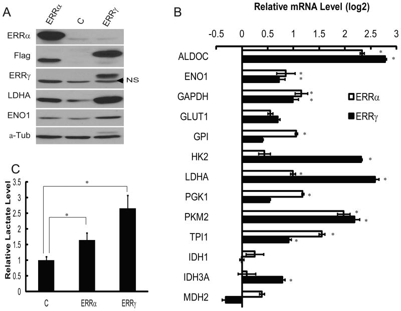
Figure 4. Overexpression of exogenous ERRs increases glycolytic gene expression and aerobic glycolysis in cancer cells.
(A) Immunoblotting analysis of ERRs and glycolytic enzymes ENO1 and LDHA in MCF7 cells transduced with control (“C”), or Flag-ERR-expressing lentivirus. Tubulin serves as a loading control. The ERRγ antibody only recognized exogenous protein. Arrowhead denotes a non-specific band (NS).
(B) Quantitative determination of glycolytic gene expression by RT-PCR in control, ERRα- and ERRγ-overexpressed MCF7 cells. * represents significant changes (p<0.05).
(C) Comparison of lactate levels in culture media from control, ERRα- and ERRγ-expressing MCF7 cells. * represents significant changes (p<0.05).

