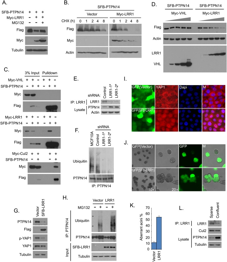
Figure 6.
The CRL2LRR1 complex is the E3 ligase that promotes PTPN14 degradation. (A) PTPN14 was degraded by CRL2LRR1. Constructs encoding SFB-tagged PTPN14 were transfected into 293T cells with or without constructs encoding myc-tagged LRR1. The indicated cells were treated with MG132 (10 μM) for 4 h. Immunoblotting analysis of the indicated proteins is shown. (B) The PTPN14 turnover rate was increased by LRR1 expression. 293T cells expressing PTPN14 with or without LRR1 were treated with cycloheximide (CHX; 100 μg/mL) for the indicated hours. Immunoblotting analysis of the indicated proteins is shown. (C) PTPN14 interacts with the CRL2LRR1 complex. 293T cells were transfected with constructs encoding SFB-tagged PTPN14 along with constructs encoding myc-tagged VHL, LRR1, or Cul2. Precipitation was carried out using S-protein beads, and immunoblotting was performed with antibodies recognizing the indicated epitope tags. (D) CRL2LRR1 targets PTPN14 for degradation. PTPN14 was expressed in 293T cells with an increased amount of VHL or LRR1. Immunoblotting analysis of the indicated proteins is shown. (E) PTPN14 expression level was increased by LRR1 knockdown. PTPN14 protein level was detected in MCF10A cells expressing the indicated shRNAs. (F) PTPN14 polyubiquitination decreased in LRR1-depleted MCF10A cells. The indicated shRNA-transduced MCF10A cells were pretreated with MG132 (10 μM) for 4 h. Immunoprecipitation was carried out using purified anti-PTPN14 antibody, and immunoblotting was performed with the indicated antibodies. (G) PTPN14 expression was decreased by the overexpression of LRR1 in MCF10A cells. Immunoblotting analysis is shown for the indicated proteins in control cells or MCF10A cells expressing LRR1. (H) PTPN14 polyubiquitination was increased by LRR1 overexpression. Control or MCF10A cells overexpressing LRR1 were cultured with or without MG132 (10 μM) for 4 h. Immunoprecipitation was carried out using purified anti-PTPN14 antibody, and immunoblotting was performed with the indicated antibodies. (I) YAP1 translocation was inhibited by LRR1 overexpression in confluent cells. Translocation of YAP1 was shown by immunostaining with anti-YAP1 antibody in confluent MCF10A cells with or without LRR1 overexpression. LRR1-positive cells are indicated by GFP expression (green). Nuclei were stained by DAPI. (M) Merged. (J) Normal polarized acini formation was disrupted by LRR1 overexpression. MCF10A cells with or without LRR1 overexpression were analyzed for acini formation in 3D culture. LRR1-positive cells are indicated by GFP expression (green). (K) Aberrant acini in MCF10A cells expressing vector or LRR1 were quantified. Data are presented as mean ± SD from three different experiments. (L) The LRR1 protein level was controlled by cell density. Immunoblotting analysis of the indicated proteins in sparse and confluent MCF10As is shown.

