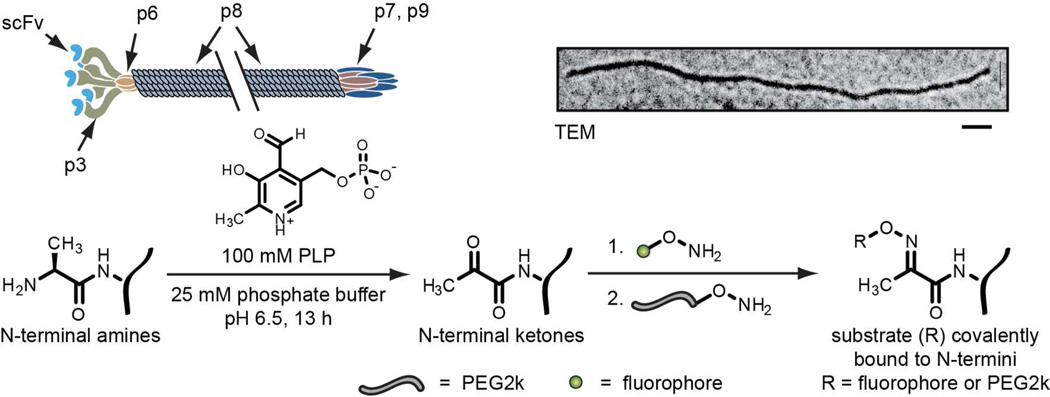
Figure 1.
A cartoon (above) and chemical scheme (below) for the transamination of filamentous phage and the attachment of synthetic molecules. The N-termini are transaminated to yield ketone-bearing proteins, which are then reacted with aminooxy-functionalized fluorophores (green circles), followed by aminooxy-functionalized PEG2k (grey strands). The double slash indicates that the phage is much longer than shown when scaled to the minor coat proteins. The TEM image was stained with uranyl acetate (top right, scale bar represents 100 nm).

