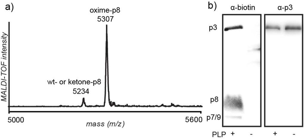
Figure 2.
Analysis of filamentous phage modified with small molecules. a) Matrix-assisted laser desorption/ionization time-of-flight (MALDI-TOF) spectrum showing p8 oxime formation following reaction with 2-(aminooxy)acetic acid (expected mass increase: 73 m/z, observed: 73 m/z). Non-transaminated fd proteins exposed to the same alkoxyamine resulted in no oxime product formation (see Supporting Information Figure S1). b) Western blot of M13KE coat protein labeling with biotin followed by blotting with neutravidin-HRP or α-p3 antibodies. Coat protein molecular weights are as follows: p3, 46.5 kD; p6, 12.4 kD; p7, 3.6 kD; p8, 5.2 kD; p9, 3.7 kD. Labeling of p7 and p9 cannot be distinguished due to their similar molecular weights (3.6 and 3.7 kD, respectively). p6 is not observed, congruent with an N-terminus inaccessible for modification.

