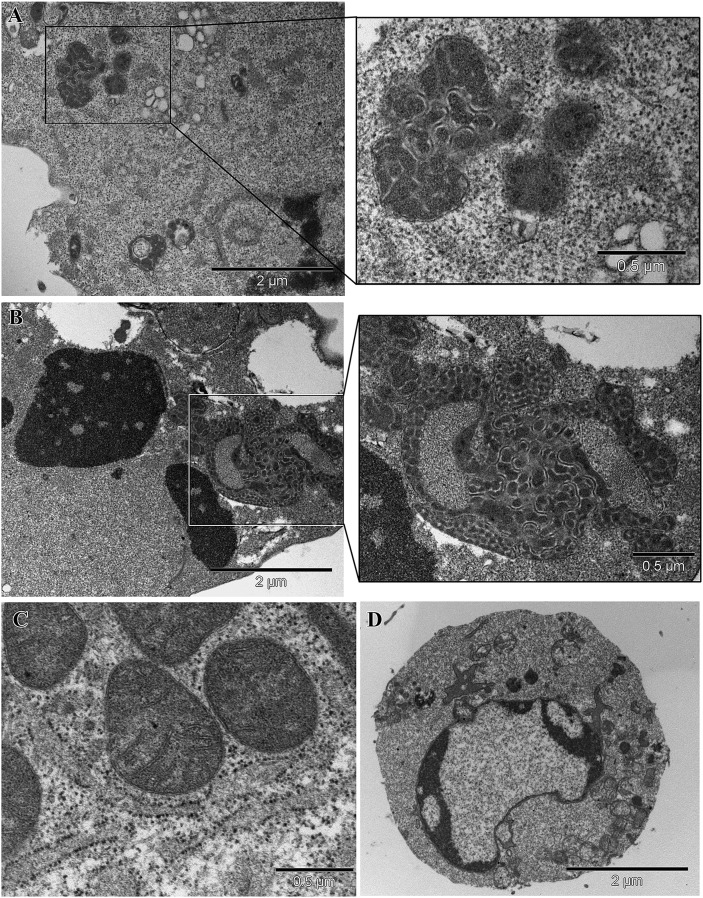
Fig. 2.
Mitochondrial features. A: At the ultrastructural level, mitochondria from HD cells appeared as electron-dense, coalescent organelles forming aggregates at one pole of the cell (inset and upper right panel). B: After treatment with STS, cells from HD patients showed chromatin aggregation typical of apoptosis as well as bolstered mitochondrial structural alterations characterized by the formation of large bundles (inset and middle right panel). C: Normal mitochondria from HS lymphoid cells. D: Control lymphoid cell undergoing apoptosis.

