Fig. 3.
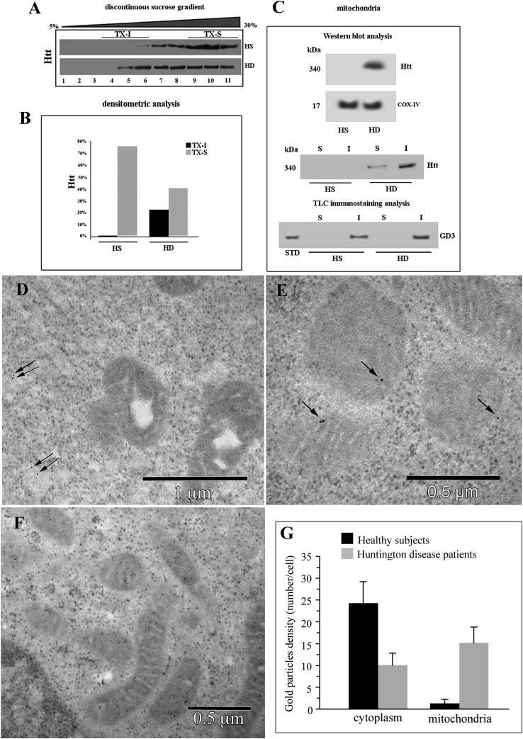
Localization of Htt at mitochondrial raft-like microdomains. A: Western blot analysis of sucrose gradient fractions. Cells from HD patients or from HS were lysed, and the supernatant fraction was subjected to sucrose density gradient. After centrifugation, the gradient was fractioned, and each gradient fraction was recovered and analyzed by Western blot using an anti-Htt polyclonal antibody. B: Densitometric analysis of sucrose gradient fractions. The columns indicate the percent distribution across the gel of fractions 4, 5, and 6 (buoyant low-density TX-100-resistant fractions, TX-I) and 9, 10, and 11 (TX-100-soluble fractions, TX-S), as detected by densitometric scanning analysis. C: Mitochondria from HD or HS cells were analyzed by Western blot and probed with anti-Htt polyclonal Ab or with anti-COX-IV MoAb. Mitochondria from HD or HS cells were solubilized by detergent as reported in Materials and Methods. Both Triton X-100-soluble (S) and -insoluble (I) fractions were analyzed by Western blot and probed with anti-Htt polyclonal Ab. Alternatively, Triton X-100-soluble (S) and -insoluble (I) fractions from mitochondria were subjected to ganglioside extraction. The extracts were run on high-performance TLC aluminum-backed silica gel and were analyzed for the presence of GD3 by TLC immunostaining analysis, using an anti-GD3 MoAb (GMR19). D–F: Transmission immunoelectron micrographs (without uranyl acetate-lead citrate counterstaining). In cells from HS, gold particles labeling Htt were very few and dispersed in the cytoplasm (arrows in D). In cells from HD patients, gold particles were detectable at the mitochondrial level (arrows in E). A representative micrograph of negative control samples stained only with gold-conjugated secondary antibody is shown in F. Results of morphometric evaluations of gold particles detected in the cytoplasm or associated with mitochondria are shown in G. Note the significant difference in the presence of cytoplasmic and mitochondria-associated gold particles between lymphoblastoid cells from HS and HD patients.

