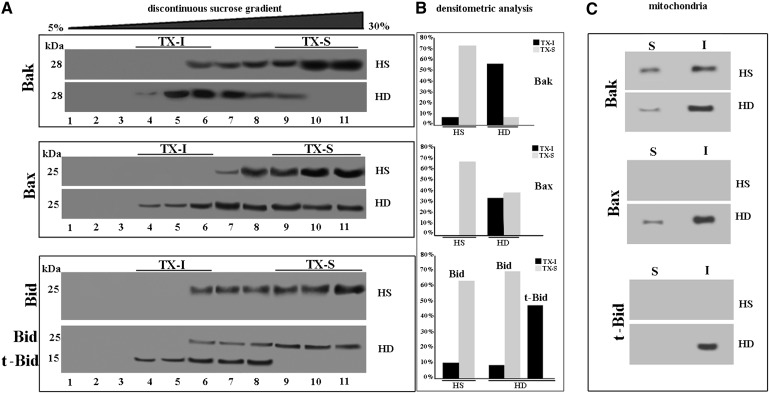
Fig. 4.
Bcl-2 family pro-apoptotic proteins. A: Western blot analysis of sucrose gradient fractions. Cells from HD patients or HS were lysed, and the supernatant fraction was subjected to sucrose density gradient. After centrifugation, the gradient was fractioned and each gradient fraction was recovered and analyzed by Western blot. First line: fractions obtained after sucrose density gradient, either HS or HD cells, were analyzed using an anti-Bak polyclonal Ab. Second line: fractions obtained after sucrose density gradient, either HS or HD cells, were analyzed using an anti-Bax monoclonal Ab. Third line: fractions obtained after sucrose density gradient, either HS or HD cells, were analyzed using an anti-Bid polyclonal Ab. B: Densitometric analysis of sucrose gradient fractions. The columns indicate the percent distribution across the gel of fractions 4, 5, and 6 (buoyant low-density TX-100-resistant fractions, TX-I) and 9, 10, and 11 (TX-100-soluble fractions, TX-S), as detected by densitometric scanning analysis. C: Mitochondria from HD or HS cells were detergent solubilized as reported in Materials and Methods. Both Triton X-100-soluble (S) and -insoluble (I) fractions were analyzed by Western blot and probed with anti-Bak polyclonal Ab, anti-Bax MoAb, or anti-Bid polyclonal Ab. The fraction samples were loaded by volume.

