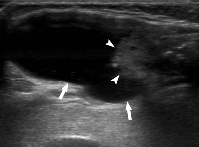
Fig. 4.
Longitudinal ultrasound image of predominantly cystic nodule in 66-year-old woman shows eccentric configuration. Note difference between smooth margin of entire nodule (arrows) and non-smooth margin of internal solid portion (arrowheads). This lesion was surgically confirmed as papillary thyroid carcinoma despite substantial cystic portion.

