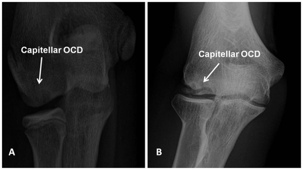
Figure 1.

(A) Anterior-posterior view radiograph of the elbow demonstrating cystic changes in the capitellum (Minami type 1) suggestive of an osteochondritis dissecans lesion. (B) Anterior-posterior view radiograph of the elbow demonstrating fragmentation and splitting of the subchondral bone in the capitellum (Minami type 2) indicative of an osteochondritis dissecans lesion.
